Digital Pathology tools
for research workflows
Advances in cancer research increasingly depend on quantitative histology and computational analysis. From whole-slide imaging to spatial tissue modeling, robust digital workflows are essential. I develop practical tools that bridge image processing and AI-driven infrastructure for reproducible biomedical research.
Featured tools
HPA Image Downloader
Automates downloading IHC cancer images from Human Protein Atlas and generates structured folders + CSV metadata summaries.
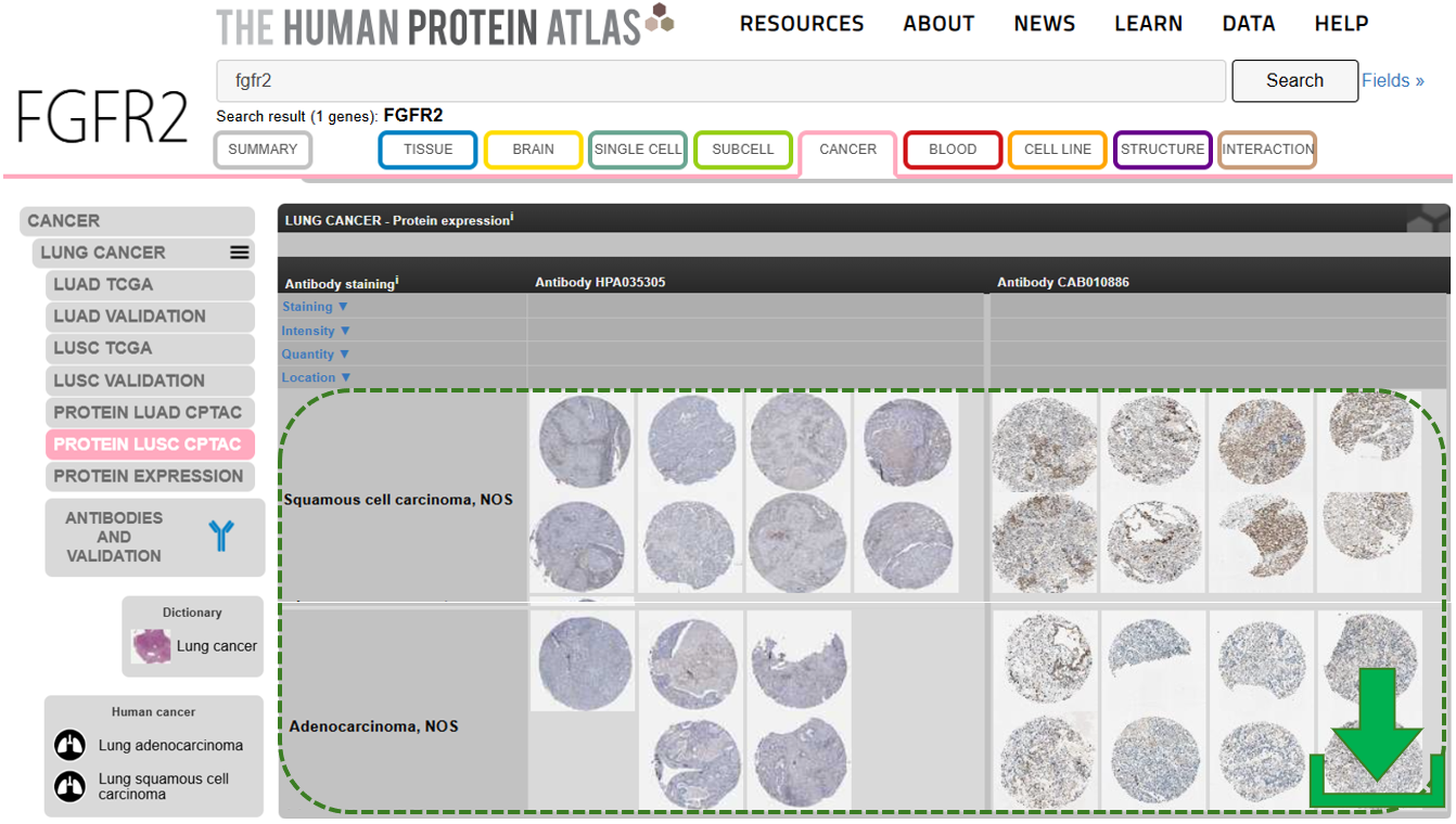
Open page
TiffCropper
Standalone Windows app to extract high-resolution ROIs from large TIFF microscopy images while preserving calibration metadata.
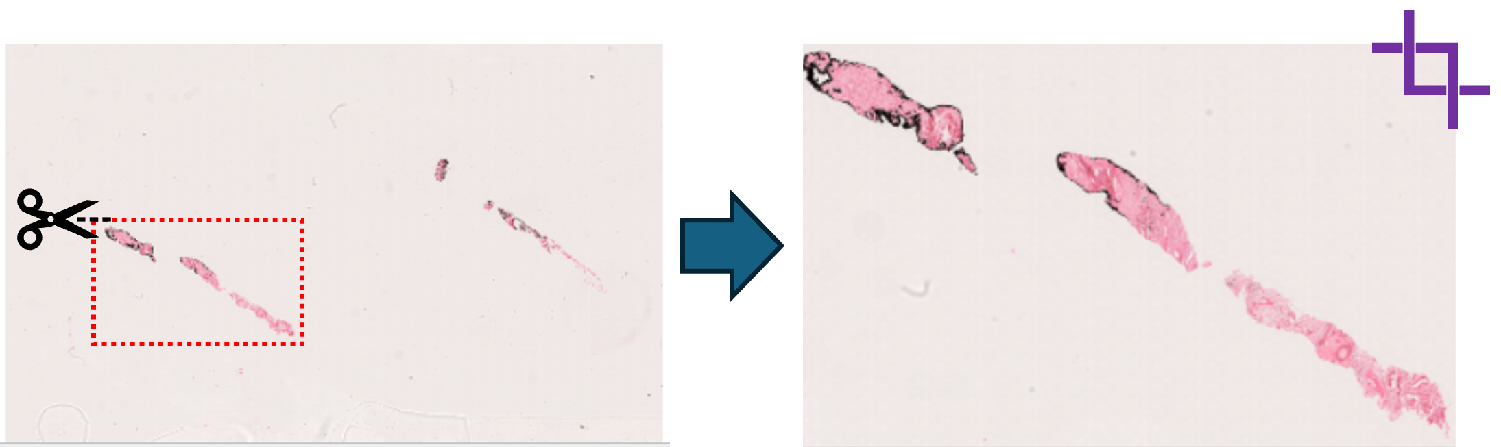
Open page
HistRegGUI
Desktop GUI for histology image registration using DeeperHistReg presets (initial, rigid, nonrigid) with CPU-only execution.
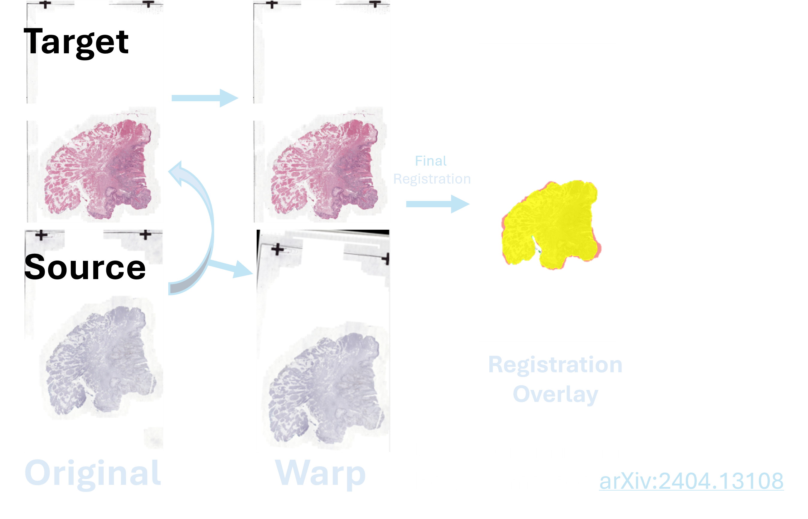
Open page
What I’m building next
Currently developing a robust end-to-end pipeline that transforms whole-slide images (WSI) into structured, analysis-ready data. The workflow includes automated tissue detection, artifact removal, efficient patch extraction and storage, and segmentation modules designed for quantitative characterization of the tumor microenvironment (TME). The goal is to enable scalable, reproducible analysis from raw histology to biological insight.