Tools
A collection of software tools designed to support scalable digital pathology workflows, from whole-slide image processing to dataset preparation, image registration, segmentation, annotation conversion and quantitative analysis.
HPA Image Downloader
Automated downloader for IHC cancer images from the Human Protein Atlas, generating structured folders and CSV metadata summaries for dataset construction.
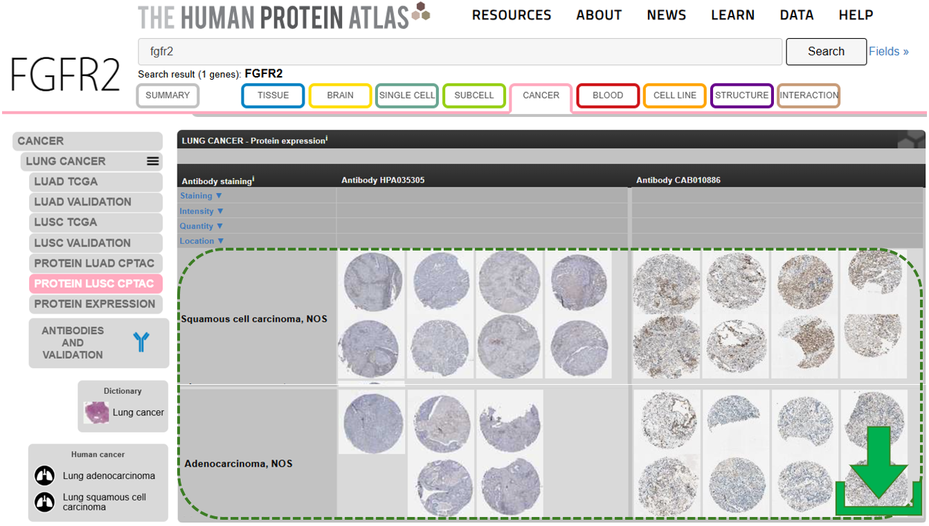
- Batch IHC image retrieval
- Structured dataset organization
- CSV metadata export
TiffCropper
Standalone Windows application for extracting high-resolution ROIs from large TIFF microscopy images, preserving calibration metadata and spatial integrity.
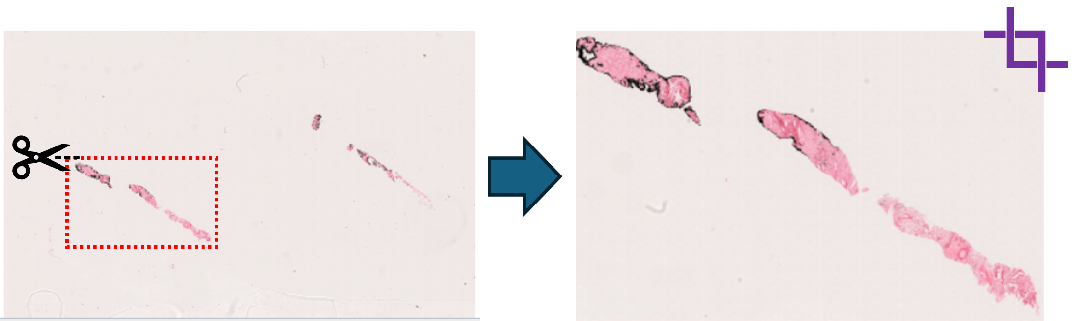
- Handles large TIFF / WSI-like images
- Metadata-safe cropping
- Designed for reproducible ROI workflows
iSyntaxToTIFF
Standalone converter for Philips .isyntax whole-slide images to pyramidal RGB OME-TIFF,
using OpenPhi and the Philips Pathology SDK.
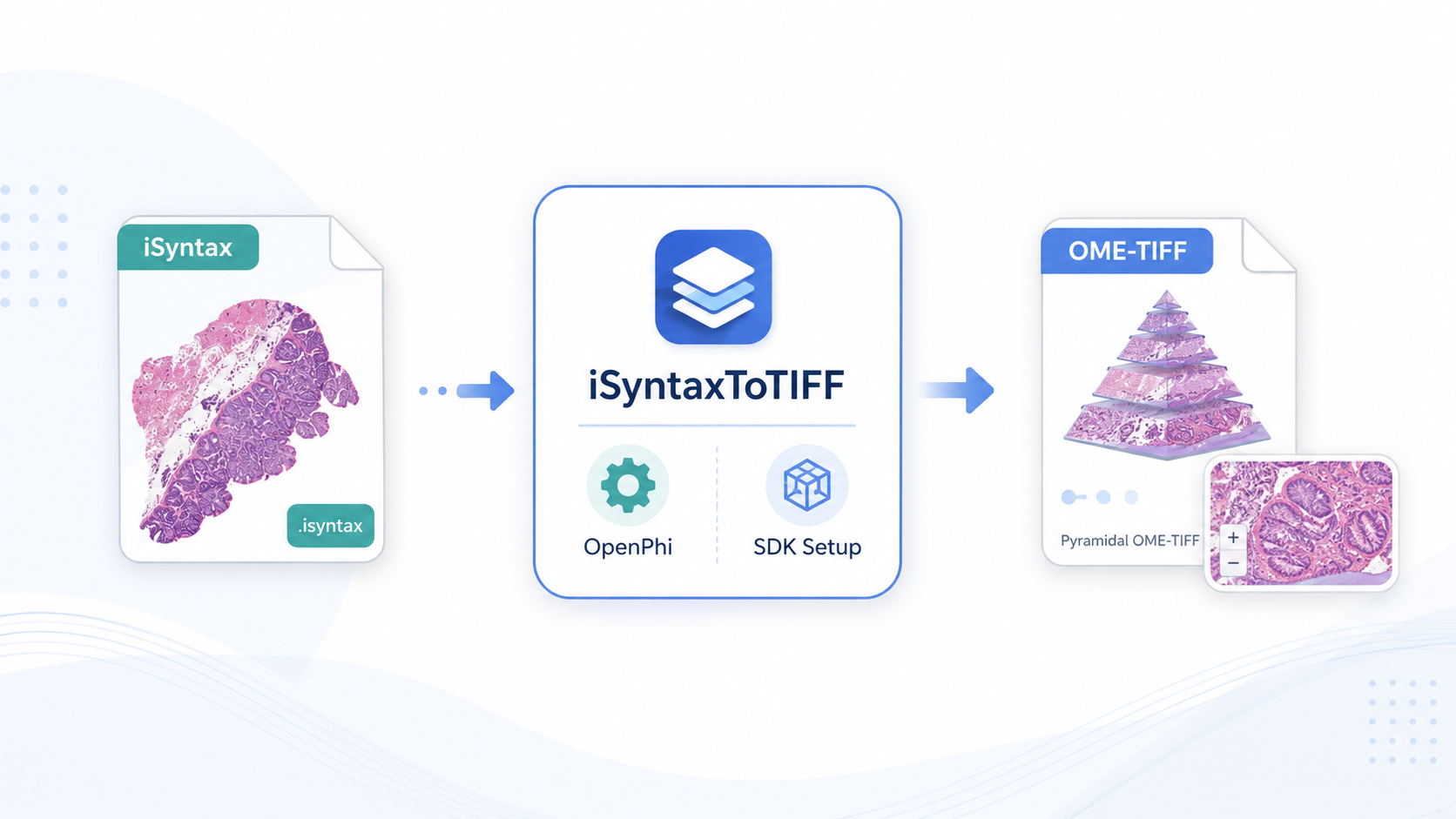
- Philips
.isyntaxto pyramidal RGB.ome.tif - SDK setup and SDK import test from the GUI
- Batch conversion with CSV conversion log
HistRegGUI
Desktop GUI for histology image registration using DeeperHistReg presets with CPU-only execution support.
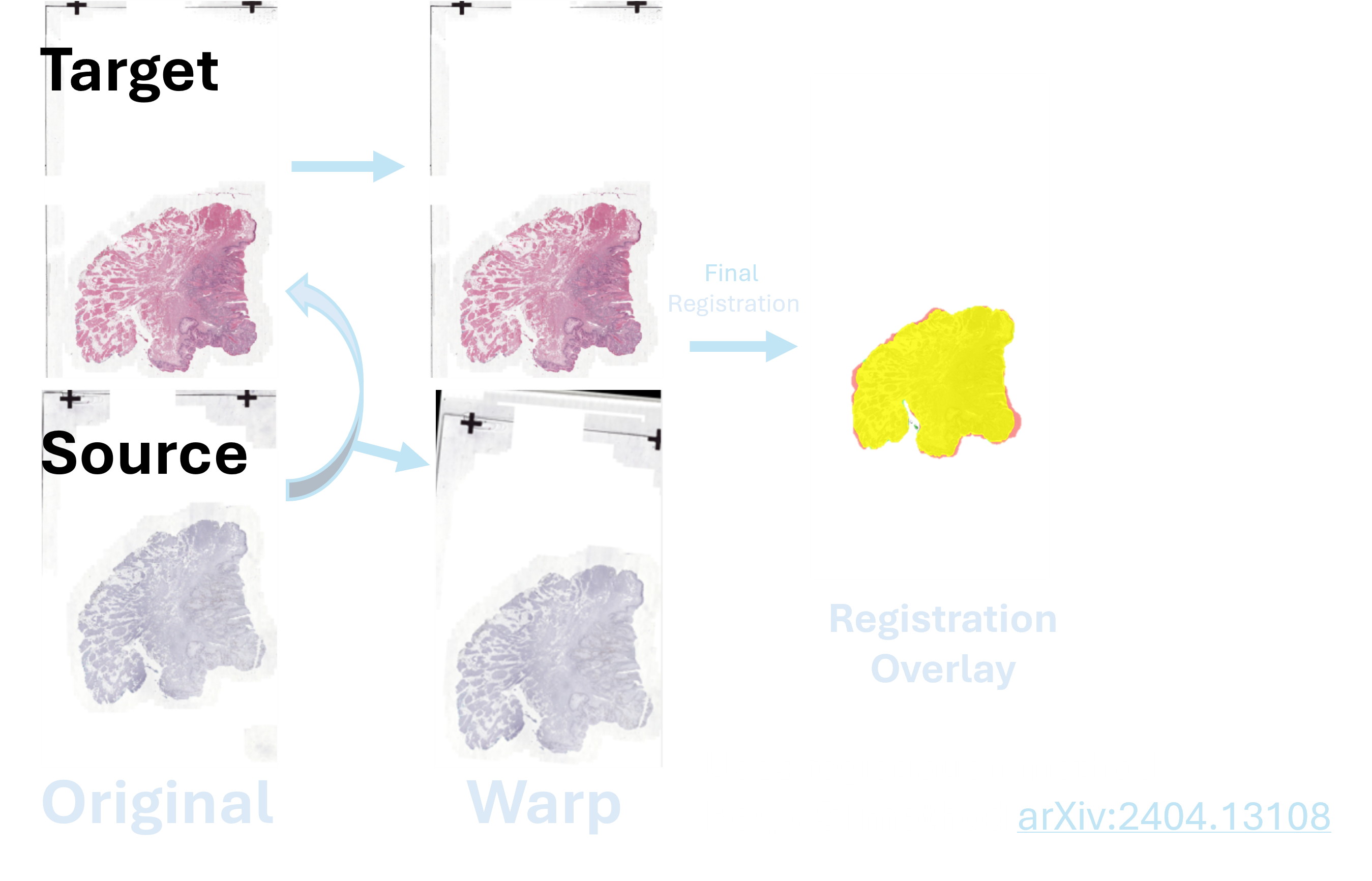
- Multi-stage registration pipeline
- CPU-friendly execution
- Designed for section alignment & 3D reconstruction workflows
Cell Well Segmentation
Desktop GUI for immunofluorescence cell segmentation, feature extraction, Manders colocalization analysis, QuPath-compatible GeoJSON export and optional DICE validation using ground-truth annotations.
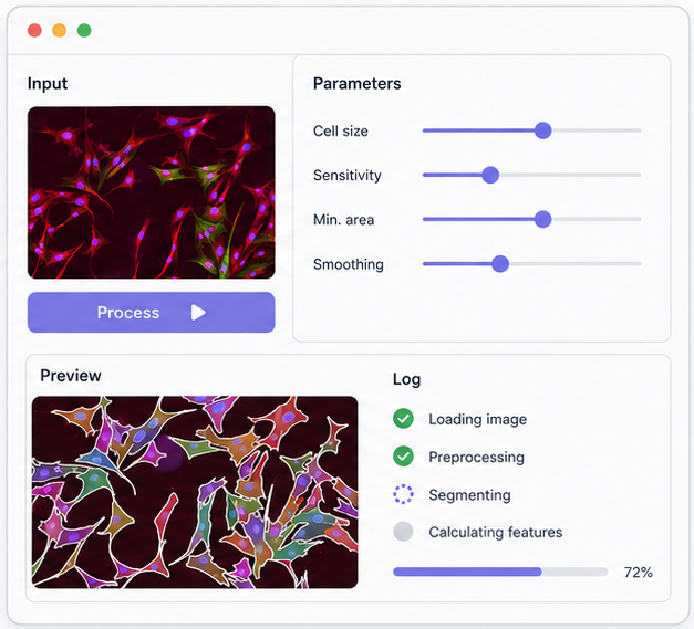
- ROI-based parameter exploration
- Instance mask, CSV, Manders and GeoJSON outputs
- DICE/IoU validation against full-image GeoJSON annotations
FeatureStat Studio
Desktop GUI for grouped statistical analysis, publication-ready plots, histograms, ROC biomarker evaluation and multi-feature batch visualization.
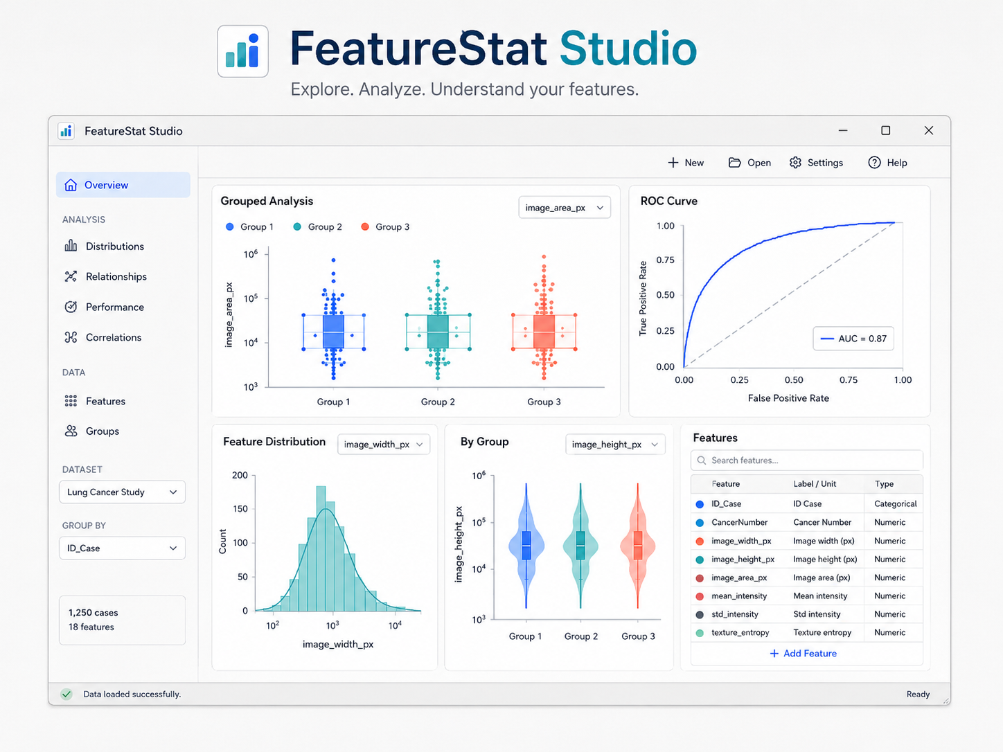
- Grouped statistics and publication-ready plots
- Histogram and ROC biomarker analysis
- Multi-feature batch preview and export
QuPath GeoJSON Converter
Desktop GUI for converting external GeoJSON annotations into QuPath-compatible annotations, detections and class-based objects.
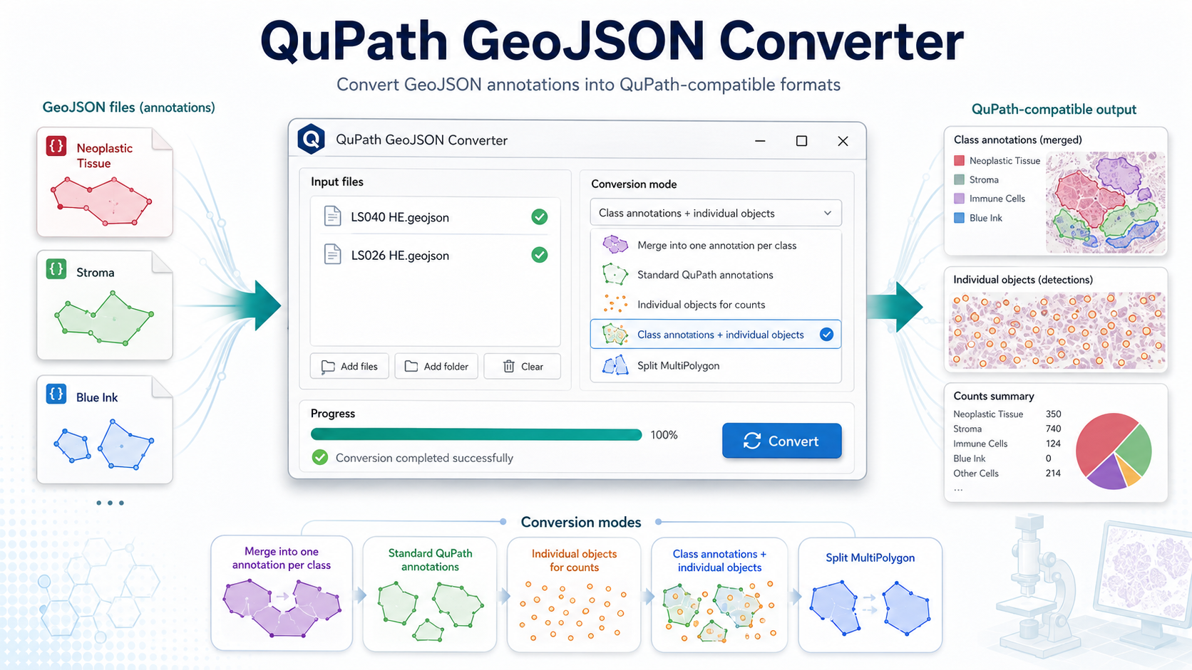
- GeoJSON classification conversion
- Annotation, detection and object-count modes
- Batch file and folder processing
Pipeline Development
Beyond standalone tools, current development focuses on an end-to-end WSI processing pipeline that transforms raw whole-slide images into structured, analysis-ready datasets.
The system integrates automated tissue detection, artifact cleaning, efficient patch extraction and storage, and segmentation modules for quantitative tumor microenvironment characterization.